The Normal Heart
The heart is a “clenched-fist” sized muscle, whose job it is to pump blood through the body 24 hours a day, 7 days a week, without rest. The heart is shaped to form four cardiac chambers. Each of the two upper chambers is called an atrium, and each lower chamber is called a ventricle. Each chamber of the heart has its own function. “Used up” blood from all parts of the body travels from the right atrium through the tricuspid valve into the right ventricle. This blood is then pumped from the right ventricle through the pulmonic valve into the pulmonary artery, which delivers the blood to the lungs to be oxygenated. The newly oxygenated blood travels from the lungs through the pulmonary veins into the left atrium. The oxygenated blood is pumped from the left atrium, through the mitral valve, into the left ventricle. Finally, during contraction of the left ventricle, the oxygen-rich blood is pumped through the aortic valve into the aorta and branching arteries to be delivered to all the organs of the body, including the brain, heart, kidneys, and muscles, only to repeat the cycle again. This coordinated action occurs because the heart is “wired” to send electrical signals that tell the chambers of the heart when to contract.
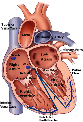 Figure 1. Anatomical view of the normal heart and conduction system. The top chambers are known as the atria. The bottom two chambers, are the left and right ventricles. A specialized network of conduction tissue exists, allowing the heart’s own pacemaker residing in the sinoatrial, or SA, node to conduct to the ventricles in a coordinated fashion.
The heart has its own electrical system, or wiring, that allows for this coordinated contractions of the heart chambers. The dominant pacemaker of the normal heart that initiates each heartbeat, is called the sinus, or sinoatrial (SA) node, and is located in the upper portion of the right atrium. The SA node typically fires at a rate exquisitely sensitive to the metabolic needs of the body, and responds to wide swings of activity, from sleeping to exercising, to ensure enough blood is directed to the body’s tissues. The electrical pacemaker signal from the right atrium spreads to the left atrium allowing coordinated contraction of the upper two chambers. Next, the electrical impulse reaches the middle of the heart, near the junction of all four chambers, at a critical structure called the atrioventricular (AV) node. The AV node, itself a natural slower pacemaker, acts like a traffic signal, regulating the flow of electrical “traffic” from the atria to the ventricles. Once the electrical impulse is allowed to pass, it reaches the His bundle and bifurcates to the branches serving the lower right and left ventricles, that is the right and left bundle branches, respectively. Specialized conduction fibers, termed Purkinje fibers, act to speedily conduct the impulse to the ventricular muscle. This synchronized sequence of atrioventricular contraction occurring under the influence of the sinus node and using the heart’s normal electrical circuit is termed sinus rhythm. Sinus rhythm provides for atrioventricular (AV) synchrony that maximizes the amount of blood pumped per each cardiac cycle. AV synchrony means that the ventricles only pump blood after they have been maximally filled via the coordinated atrial contractions. This AV synchrony optimizes cardiac output and produces the greatest efficiency of the cardiovascular system.
 Figure 2. Animation of normal sinus rhythm. The dominant pacemaker, the sinus node, fires with electrical depolarization of the atrium spreading through the AV node to the bottom ventricles, allowing for AV synchronous contraction.

 Silver Spring Office
  DC Office (at Providence Hospital)
  Hagerstown Office