Atrial Flutter
Overview
Atrial flutter, another type of supraventricular tachycardia, produces a heart rhythm with typically more atrial contractions than ventricular beats. The atria can contract anywhere from 250-400 beats per minute, with the AV node serving as the “traffic control” preventing 1 to 1 conduction to the ventricles. The heartbeat on exam is typically fast, but with medications, it can be slow with either a regular or irregular pulse.
Atrial flutter is the result of a rapid electrical circuit localized to the top right heart chamber, the right atrium. This electrical circuit has been extensively mapped using electrophysiology-guided catheter mapping techniques. It can occur in patients with no structural heart disease (lone atrial flutter), but is more likely in those with a history of prior cardiac surgery, congestive heart failure, or congenital heart disease. Conditions that lead to right-sided heart dilation and increased pressure can increase the incidence of this rhythm disturbance (right-sided heart failure, pulmonary hypertension, COPD, hypertension, obesity, tricuspid or pulmonary stenosis).
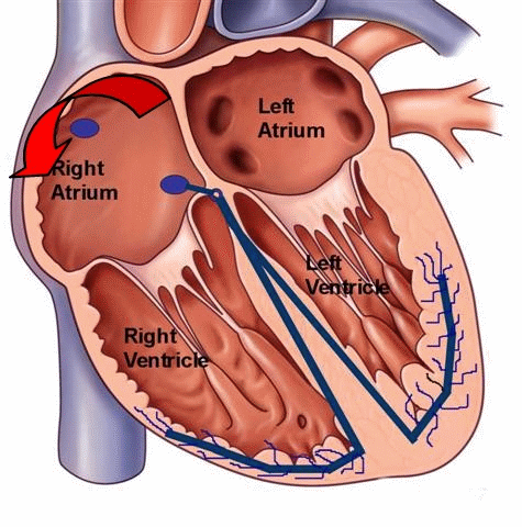
Figure 1. Animated illustration of the electrical circuit in the right atrium during typical atrial flutter. Ablation of this rhythm targets a critical portion of the circuit know as the cavotricuspid isthmus (CTI); it is to this area bounded by the tricuspid valve and the inferior vena cava (IVC) that a linear ablation lesion can be delivered with successful interruption and termination of the circuit.
Symptoms
Episodes of atrial flutter can lead to rapid heartbeats with symptoms of palpitations and chest fluttering or tremoring. Similar to other SVTs, patient may feel short of breath, fatigue, dizziness, or rarely, even fainting. However, some people with atrial flutter may not have any symptoms at all. Episodes of atrial flutter can last anywhere from minutes to days or months. Patients with sustained rapid heartbeats should seek medical attention.
Diagnosis
Atrial flutter can be diagnosed by your physician via an electrocardiogram or an Ambulatory monitoring device, i.e. Holter or Event monitor, specifically during an arrhythmia episode. Some episodes of atrial flutter are intermittent (also referred to as paroxysmal) and not reliably present on a daily basis. In these situations, your physician may recommend a special ambulatory monitoring device that is patient-triggered, i.e. an Event monitor.

Figure 2. EKG strip of atrial flutter with the “flutter waves” denoted by the red arrows. The ventricular response, or heart rate, is determined by the ratio of atrial beats that are conducted to the ventricles via the normal AV node. It appears to be 3:1, with every 3 atrial flutter waves associated with one ventricular beat.
Prognosis
Atrial flutter, with appropriate treatment, is rarely life-threatening. However, similar to atrial fibrillation, atrial flutter poses an increased risk of stroke. This is a direct results of the rapid atrialcontraction that leads to a stasis or pooling of blood in the atria; particularly, the left atrium. This pooled blood is more likely to form clots that can travel to the systemic circulation and the brain, leading to a cerebrovascular accident or stroke. The risk of stroke is not the same for all people with atrial flutter. Therefore, consultation with your physician is very important in determining the presence of additional risk factors for stroke that may require treatment with the blood thinner warfarin (also known as coumadin). Some of these risk factors include age, presence of structural heart disease, hypertension, diabetes, history of stroke, valvular heart disease, heart failure, and coronary artery disease. The decision to implement long-term anticoagulation (that is blood thinners) must be individualized after a discussion between you and your treating physician.
In addition to the risk of stroke, prolonged episodes of atrial flutter can cause irreversible changes to the atria, including negative remodeling with atrial enlargement and weakness (myopathy). In addition, prolonged episodes can make reversion and maintenance of normal sinus rhythm more difficult. Long-lasting atrial flutter with continuously rapid ventricular rates (heart rates typically greater than 100 beats per minute) may cause a tachycardia-induced cardiomyopathy (weakening of ventricular muscle and heart function) and symptoms of congestive heart failure. Atrial fibrillation and atrial flutter tend to coexist, with the significance of atrial flutter in the initiation and perpetuation of atrial fibrillation recently becoming more apparent. It is not uncommon to find patients who have frequent paroxysms of atrial fibrillation post radiofrequency catheter ablation ofatrial flutter. Eradication of atrial flutter may or may not improve the burden of atrial fibrillation in these patients. In addition, patients with atrial fibrillation who are treated with antiarrhythmic medications or post catheter ablation with pulmonary vein isolation are noted to revert to atrial flutter. In these situations, atrial flutter can be approached with a catheter ablation procedure (discussed below), with eradication of the patient’s atrial tachyarrhythmias.
Therapy
Therapy for patients suffering from atrial flutter has three components:
- Control of the ventricular rate (heart rate)
- Conversion and maintenance of sinus rhythm
- Reduction of subsequent stroke risk (as discussed earlier, with anticoagulation)
 Medications used to control the ventricular rate during atrial flutter include b-blockers, calcium channel blockers, and less commonly digoxin. These medications can be administered orally on a routine outpatient basis, or via intravenous route if necessary in the emergency room. Some patients may need a combination of medications from different classes. Patients may experience alleviation of symptoms due to slowing of the ventricular response (heart rate) with the use of b-blockers or calcium-channel blockers that delay electrical conduction through the AV node.
Attempts to convert and maintain sinus rhythm can be approached in one of three ways:
- In-hospital electrical or chemical cardioversion,
- Outpatient antiarrhythmic medicationtherapy, or
- Catheter ablation
Electrical cardioversion is a procedure done typically in the hospital, where the patient is brought to a specialized room and briefly sedated to sleep. External defibrillation pads are placed in defined locations on the patient’s chest, with the delivery a brief electric shock (less than a second). The success rate of converting atrial flutter to normal sinus rhythm is very high with this procedure. However, it does not guarantee freedom from future reversion back to atrial flutter. Chemical cardioversion can also be performed with the infusion of intravenous antiarrhythmic medications. The success rate of this procedure, in comparison to external electrical cardioversion, can be lower especially in patients with prolonged episodes of atrial flutter. In addition, some intravenousantiarrhythmics can be contraindicated in subsets of patients with structural heart disease, thus limiting their applicability. Electrical or chemical cardioversion in a patient with a prolonged episode of atrial flutter (>48 hrs) can only be performed after 4 weeks of documented appropriately therapeutic INRs (measures level of blood anticoagulation) on outpatient warfarin therapy. Treatment with anticoagulation for this 4 week time-frame has been shown to reduce, but not eliminate, the risk of stroke with the cardioversion procedure, by preventing the formation of additional clots in the left atrium. Alternatively, if a cardioversion is more urgent, or if a patient wishes to undergo a more immediate catheter ablation procedure, a transesophageal echocardiogram can be performed to screen for the presence of clots in the left atrial appendage the day of the procedure.
Outpatient oral antiarrhythmic therapy can be instituted in those patients who are typically on oral anticoagulation therapy. They can be helpful in maintaining normal sinus rhythm and prevent recurrences of atrial flutter after electrical cardioversion. Your physician may opt to start antiarrhythmic medications in the weeks prior to your cardioversion procedure. There are many antiarrhythmic medications, with only a few requiring a brief hospital stay for initiation. These pharmacologic therapies act directly on the cardiac substrate by slowing electrical conduction in the heart and lengthening the time required for electrical recovery of heart tissue. Drug therapy for atrial flutter can be a “trial and error” proposition; it is not possible to determine ahead of time which medication will work the best in a particular patient. Maintenance of sinus rhythm with complete suppression of atrial flutter may not always be possible with medications. Improvements in your quality of life must constantly be reevaluated in relation to the small risks, discomfort, and potential side effects associated with your prescribed treatment regimen. Antiarrhythmic medications are not without risk, as they can increase the incidence of other cardiac arrhythmias.
For those patients who elect to forgo medication either due to
- intolerable symptoms or side effects from their medications,
- recurrent symptoms and episodes despite medical therapy, or
- lack of desire to take daily medications for an extended period of time,
your physician may recommend you undergo an electrophysiology study (EPS) and possible “curative” catheter ablation. In brief, these procedures are performed in a special procedure room called an electrophysiology (EP) laboratory. The EP laboratory houses a table and an x-ray (fluoroscopy) machine that is suspended over the table. With the use of specialized electronic and computer equipment, in conjunction with the intracardiac electrode catheters (placed into the heart via the femoral veins), your heart rhythm specialist can map the right atrial flutter circuit and deliver radiofrequency energy to the site, thus interrupting the circuit and terminating the tachycardia.Radiofrequency energy employed during the catheter ablation procedure heats the tissue enough to destroy the local heart cell function, but without physically cutting through the tissue. The long-term success rate is very good for atrial flutter, but recurrences can occur as the heart attempts to heal itself. These recurrences can be treated with a second catheter ablation procedure. Not every patient is a candidate for an atrial flutter catheter ablation procedure, and an extensive conversation with your physician is important to review the various treatment options with you. As alluded to earlier, atrial flutter may coexist with other atrial tachyarrhythmias, including atrial fibrillation. Catheter ablation of the atrial flutter circuit may reduce the burden of coexistent atrial fibrillation in these patients, but likely it will not allow immediate withdrawal of anticoagulation therapy in most patients.
- Arrhythmias
- Sick Sinus Syndrome
- Atrioventricular (AV) Block
- Atrial Tachycardia
- AV nodal reentrant tachycardia (AVNRT)
- Wolff-Parkinson White Syndrome (WPW) and Atrioventricular reciprocating tachycardia (AVRT)
- Atrial Flutter
- Atrial Fibrillation
- Cardioversion
- Ventricular Tachyarrhythmias
- Long QT Syndrome
- Sudden Cardiac Death (SCD)
- Syncope


 Silver Spring Office
  DC Office (at Providence Hospital)
  Hagerstown Office